


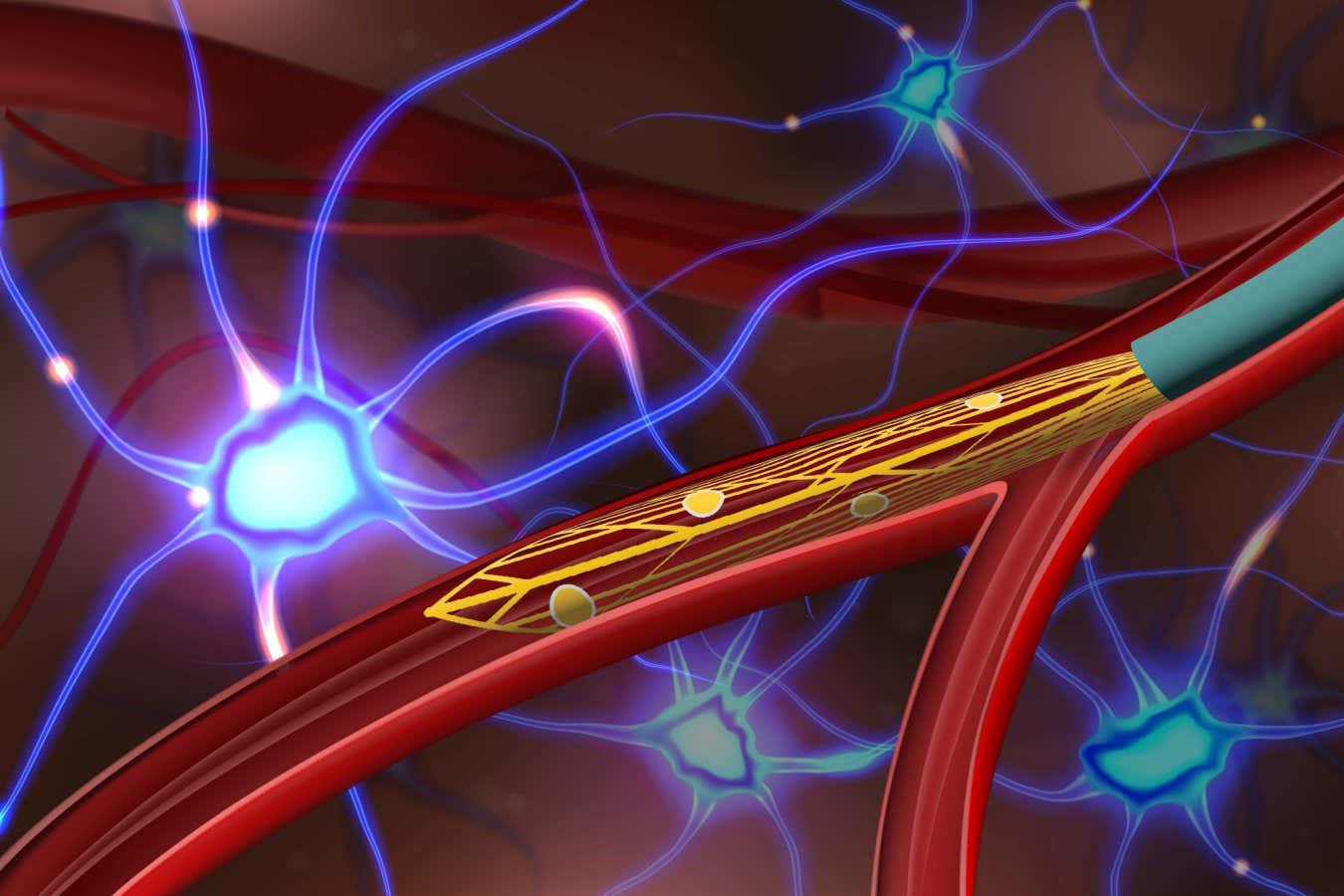
The probe (yellow) can be inserted into a blood vessel via a catheter (cyan)
Anqi Zhang, Stanford University
Introduction
In a groundbreaking development, scientists have successfully injected rats with a small, flexible probe that can record deep brain activity without the need for surgical implantation. This state-of-the-art equipment has the potential to revolutionise the field by providing a non-invasive means of diagnosing and treating a wide range of brain disorders in humans.
The development and use of brain-machine interfaces (BMIs) have been crucial to the study of the brain and the treatment of neurological illnesses like Parkinson’s disease. However, traditional BMIs require invasive procedures for accurate recording. The latest innovation holds promise in overcoming this limitation, and this article will delve into the details of this remarkable advancement.
Brain-Machine Interfaces (BMIs) and Their Significance
In order to record and transfer electrical impulses from the brain to external computers, brain-machine interfaces (BMIs) have been developed as implantable devices. These interfaces are crucial tools in studying brain functions and aiding in the treatment of neurological conditions. For instance, they have been instrumental in the management of Parkinson’s disease, providing relief to patients through deep brain stimulation.
The Challenge of Invasive BMIs
While BMIs have proven effective, they come with a trade-off between invasiveness and recording resolution. Some BMIs employ sensors on the scalp, but the recording quality is often compromised due to the skull’s interference with electrical signals. To accurately measure brain activity, surgical implantation of electrodes through open-skull surgery has been the preferred method.
The Revolutionary 7-Centimeter-Long Probe
Charles Lieber and his team at Harvard University have developed an innovative solution to the invasiveness issue. They have created a 7-centimeter-long mesh-like probe embedded with 16 electrodes, utilizing a flexible polymer. This probe can navigate through the intricate network of blood vessels in the brain, enabling it to reach deep brain regions more accurately.
Implantation Procedure in Rats
In their groundbreaking study, the researchers made a small incision in the rats’ necks and skillfully guided the probe to the base of their brains using a thin catheter. Electrodes were able to pick up signals from neurons when the probe expanded and attached to the walls of the blood vessel.
Electrode Quantity and Accuracy
While the current prototype probe contains a relatively small number of electrodes compared to traditional surgical implants, it still demonstrates high accuracy in signal recording. The researchers note that increasing the number of electrodes could further enhance accuracy, making it even more effective in monitoring brain activity and detecting conditions.
Human Trials and Potential Applications
The team’s next step is to conduct trials of this technology in humans, with the potential to revolutionize the treatment of conditions such as epilepsy and Parkinson’s disease. However, Charles Lieber emphasizes that ensuring the safety and effectiveness of the probe in humans will require time and rigorous testing.
The Vision for Minimally Invasive Brain Implants
Salman Qasim at Icahn School of Medicine at Mount Sinai in New York lauds this study’s potential in paving the way for minimally invasive brain implants within blood vessels. This could offer unparalleled access to intricate brain regions with complex arrangements of blood vessels, making the treatment of neurological disorders more targeted and effective.
Conclusion
In conclusion, the breakthrough development of a small, flexible probe for deep brain activity monitoring in rats holds significant promise for the future of brain-machine interfaces. By overcoming the challenges of invasiveness and low resolution, this innovation opens up new avenues for the treatment of neurological conditions in humans. The successful human trials of this technology will be eagerly anticipated, as it could revolutionize the field of neuroscience and enhance the lives of millions worldwide.
FAQs (Frequently Asked Questions)
Q1: How does the new probe differ from traditional brain-machine interfaces?
The new probe developed by Charles Lieber and his team offers a minimally invasive approach to record deep brain activity, eliminating the need for surgical implantation. It utilizes a flexible polymer and can navigate through blood vessels, providing more accurate recordings.
Q2: What are the potential applications of this technology in humans?
The technology has potential applications in various brain conditions, such as epilepsy and Parkinson’s disease, where accurate monitoring and targeted treatment are crucial.
Q3: How many electrodes does the current prototype probe contain?
The current prototype probe is embedded with 16 electrodes, while traditional surgical implants can have around 1000 electrodes.
Q4: When will human trials of this technology begin?
The researchers are planning to conduct human trials; however, proving the safety and effectiveness of the probe will take time and careful evaluation.
Q5: How could minimally invasive brain implants benefit patients?
Minimally invasive brain implants, as envisioned by Salman Qasim, could provide access to complex brain regions through blood vessels, leading to more effective and targeted treatments for neurological disorders.