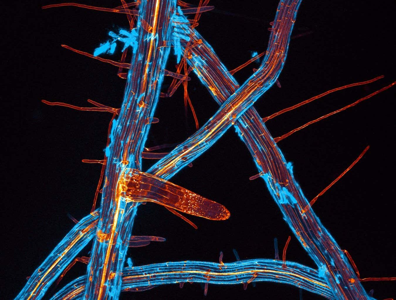
The minute details of the roots of Arabidopsis thaliana
Jan Martinek
Photographer Jan Martinek
These images of plant cells, taken by Jan Martinek, offer a vibrant and diverse perspective on plants. These images showcase the intricacy and beauty of plant cells and molecules using fluorescence microscopy. Martinek, a plant cell biologist, is not only interested in the mechanisms that shape these structures but also wants to promote science to the public by sharing such visually appealing images on his Instagram account, @plant_microverse.
A hollyhock pollen grain viewed using a confocal microscope
Jan Martinek
One of the images showcases the minute details of the roots of Arabidopsis thaliana, a small weed widely used as a model organism in genetics research. Another image captures a hollyhock pollen grain using a confocal microscope, a type of fluorescence microscopy that enhances optical resolution and contrast.
The remaining images reveal the inner “plumbing” of two plants. The first image shows the rhizome, an underground stem from which roots and shoots protrude, of a common reed. The second image depicts the hollow stalk of a barley plant, with the blue fluorescence indicating lignin, a polymer that provides the “backbone” for plant cell walls, and the red marks indicating areas of active photosynthesis.